Neurovascular Cases

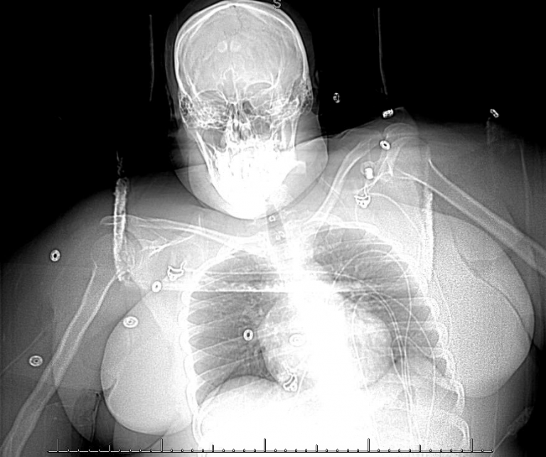
scout-ap-2

What’s the diagnosis?

The CT head shows diffuse cerebral edema or swelling and also small regions of subarachnoid hemorrhage. Injections of the bilateral internal carotid arteries reveal sluggish transit times and no normal venous outflow. The patient has profound cerebral venous sinus thrombosis.

Background

  • Patients with cerebral venous sinus thrombosis tends to either make a significant recovery or have a very poor outcome, depending on whether or not cerebral venous outflow can be restored quickly enough.
  • The traditional standard of care is anticoagulation, typically intravenous heparin.
  • Endovascular therapy may restore venous outflow more rapidly than systemic anticoagulation by physically breaking up and removing the mechanical blockage.

What's your treatment plan?

  • To reach the dural venous sinuses catheters are advanced in a retrograde approach from the femoral vein → vena cava → internal jugular vein → dural venous sinuses.
  • Within the sinuses, which have strong dural walls, aggressive thrombectomy can be performed and thrombolytic agents can be administered. In contrast, the cortical veins that drain into the dural sinuses are very fragile and manipulation of these draining veins may cause venous perforation and hemorrhage.

In this case extensive thrombectomy using manual aspiration and pump aspiration was able to partially recanalize the superior sagittal sinus, the torcula, and the right transverse sinus. Because the right sigmoid sinus would not remain patent, and because pressure monitoring demonstrated elevated intracranial pressures, the right sigmoid sinus was stented open and venous outflow from the brain through the right venous sinus system was restored.

Injection of the right jugular bulb demonstrates a patent right internal jugular vein. It was necessary to demonstrate patency of the right internal jugular vein when planning the stent construct, because the jugular vein would be the distal portion of the outflow tract.


Telescoping stents were placed in the right transverse and sigmoid dural venous sinuses, plastering residual thrombus against the sinus walls and restoring venous outflow. The stents were placed in an overlapping, or telescoping fashion.


The pressure monitoring micro-wire was advanced into the torcula and revealed that once venous outflow was restored the intracranial pressure normalized.