Neurovascular Cases

ct-head
cta
t2
t1c

What’s the diagnosis?

Tentorial dural arteriovenous fistula filling from the left middle meningeal artery and the left posterior meningeal artery with rapid cortical venous reflux into the right cerebellum.

ct-head

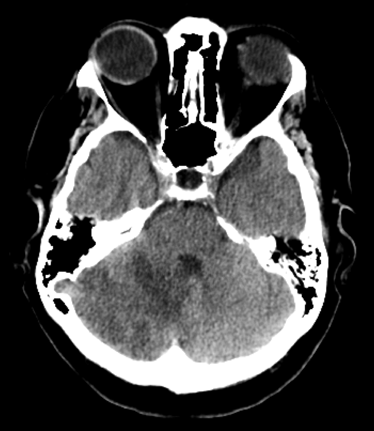
Axial CT head shows right cerebellar hypoattenuation.


cta

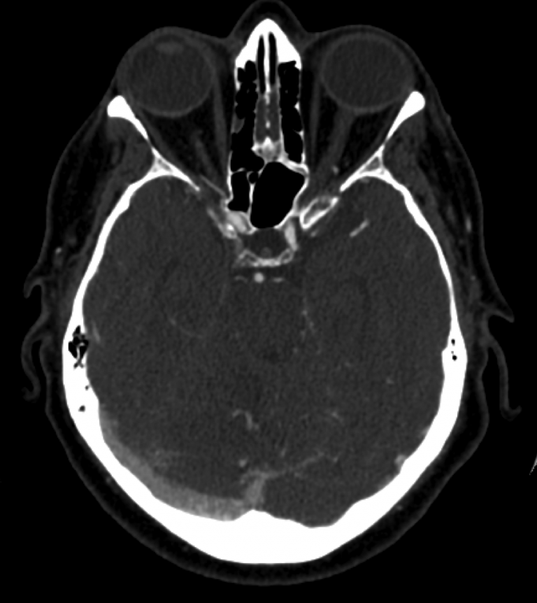
Axial CTA shows predominance of venous outflow through the right transverse sinus and minimal if any filling of the left transverse sinus.


t2

Axial T2 MR brain shows right cerebellar hyperintensity, indicating venous congestion. Prominent right cerebellar serpiginous flow voids indicate dilated veins.


t1c

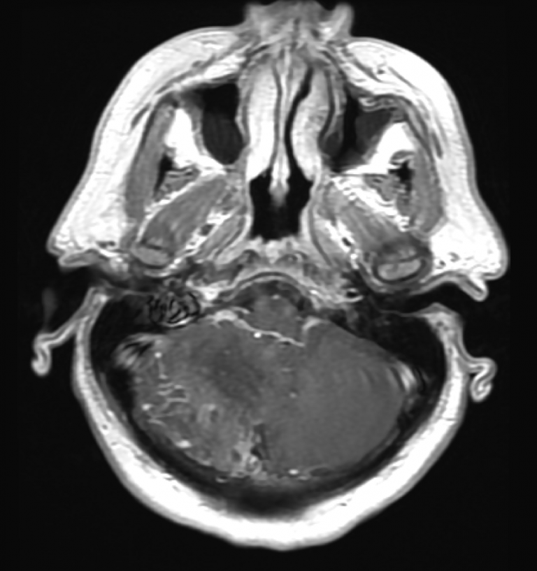
Axial T1+C MR brain shows right cerebellar focal enhancement, indicating chronic venous congestion.


Lateral projection of left external carotid artery shows a distal middle meningeal artery that supplies a dural arteriovenous fistula of the tentorium with rapid cortical venous reflux toward the cerebellum.


Lateral projection of left external carotid artery injection with focus on middle meningeal artery redemonstrates supply to the tentorial dural arteriovenous fistula with rapid cortical venous reflux toward the cerebellum.


Lateral projection of a left vertebral artery injection shows a robust posterior meningeal artery that supplies a dural arteriovenous fistula of the tentorium with rapid cortical venous reflux toward the cerebellum.


Differential Diagnosis

  1. Dural arteriovenous fistula
  2. Pial arteriovenous malformation

Background

  • Abnormal connection between arteries that would normally feed the meninges, bone, or muscles but not the brain and small venules within the dura matter
  • Account for 15% of all intracranial arteriovenous shunts
  • Lesions without cortical venous reflux are benign
  • Lesions with cortical venous reflux have an aggressive clinical course

What's your treatment plan?

  • Treatment options for dAVF include conservative therapy, endovascular embolization of the fistula, microsurgical disconnection of the fistula, and stereotactic radiosurgery
  • In this case the dAVF demonstrated significant cortical venous reflux and caused neurological symptoms
  • Given the malignant natural history of dAVFs with cortical venous reflux that present with aggressive symptoms, conservative therapy was felt to be inappropriate

In this case, endovascular transcatheter embolization of the left middle meningeal artery and the left posterior meningeal artery with N-butyl cyanoacrylate and Onyx was performed.