Neurovascular Cases

cp-t2flair-2
cp-t2flair2-2
cp-t2-coronal-2

What’s the diagnosis?

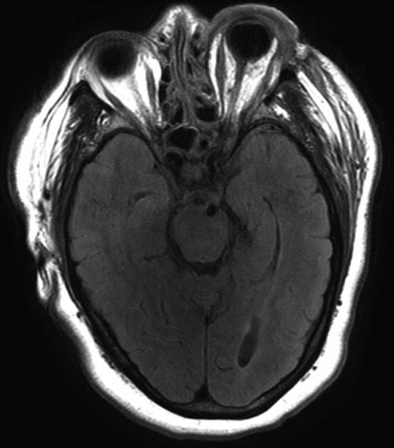
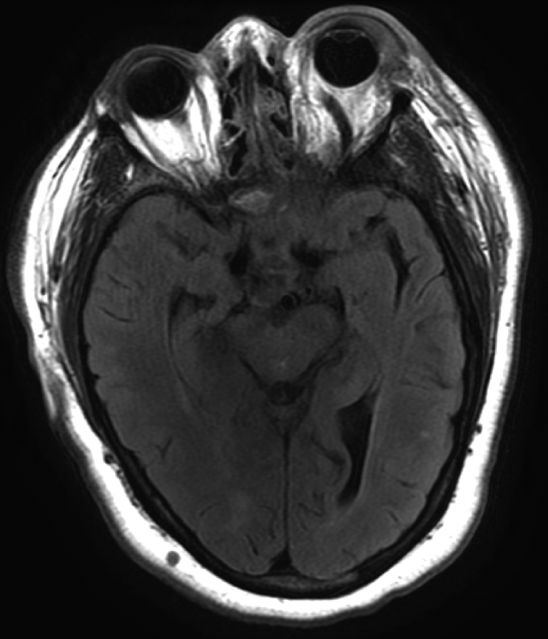
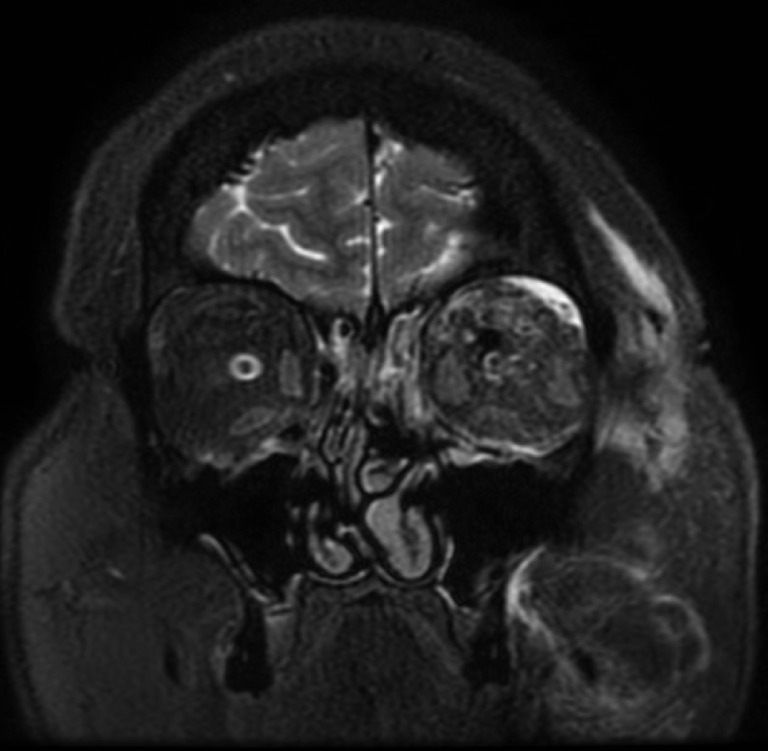
Spontaneous, left-sided direct carotid artery-cavernous sinus fistula

Left internal carotid artery injection, transfacial view demonstrates nearly immediate shunting of arterial blood into the left cavernous sinus. The left sided arterial system flow continues and ACA and MCA filling is superimposed on diffuse venous outflow routes. Venous drainage pathways are bilateral, and there is robust cross-flow from the left cavernous sinus through the intercavernous sinus to the right cavernous sinus. Venous drainage is seen through the petrosal sinuses, pterygoid plexus, and ophthalmic veins.
Background

  • A direct carotid artery-cavernous sinus fistula (CCF) means that the internal carotid artery is directly shunting high-pressure arterial blood into the normally low-pressure venous cavernous sinus.
  • Venous hypertension drives the pathophysiology of a CCF and an understanding of the venous outflow can predict what symptoms the patient may experience.
  • The classic clinical triad consists of 1) pulsatile exophthalmos, 2) orbital bruit, and 3) conjunctival injection. Because the shunt is so high flow in direct CCF, visual function is at risk.
  • While the majority of direct CCFs are traumatic, they can be spontaneous as well, such as in the case of a ruptured internal carotid artery aneurysm.

What's your treatment plan?

  • Endovascular obliteration of the fistulous connection is the first-line treatment option. However, this can be done in several ways: The internal carotid artery can be sacrificed (→ ↓ arterial inflow to the fistula); Embolization of the cavernous sinus can be performed trans-arterially (i.e., from the internal carotid artery, through the fistula, and into the cavernous sinus → ↓ potential space for arterial inflow); or Embolization of the cavernous sinus can be performed trans-venously (i.e., through some venous inlet into the cavernous sinus → ↓ potential space for arterial inflow).
  • Open microsurgical repair of the fistula is highly invasive and much less common.

In this case, transvenous embolization of the left superior ophthalmic vein and left cavernous sinus was performed via the left inferior petrosal sinus. Embolization was performed using coils. A balloon was temporarily inflated within the internal carotid artery during the procedure to ensure that coils remained in the cavernous sinus and did not prolapse into the carotid artery.