Neurovascular Cases

What’s the diagnosis?

Unruptured 6 mm left A3-segment pericallosal artery aneurysm with a wide neck and no associated branch artery origin in a patient with a history of familial aneurysms.

cb-mag-view

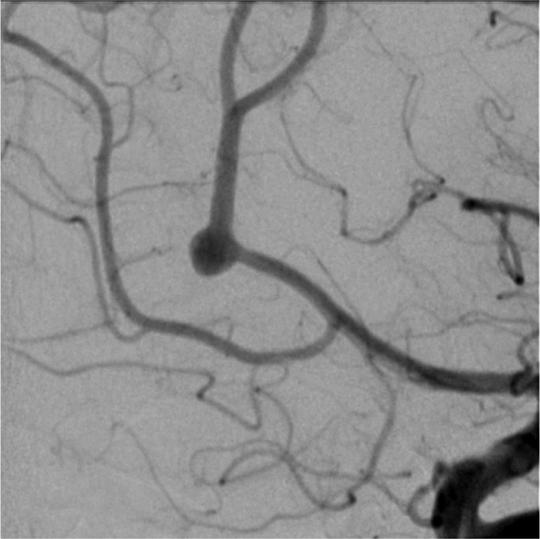
Magnified left ICA injection image, lateral view demonstrates the aneurysm in greater detail.


Left ICA injection, Townes view shows brisk filling of the anterior cerebral artery and middle cerebral artery candelabras. There is a wide necked pericallosal aneurysm that is not associated with a branch artery origin.


Left ICA injection, lateral view shows brisk filling of the anterior cerebral artery and middle cerebral artery candelabras. There is a wide necked pericallosal aneurysm that is not associated with a branch artery origin.


Differential Diagnosis

  1. Distal left anterior cerebral artery aneurysm

Background

  • The anterior cerebral artery is divided anatomically into five segments, the first of which are A1) from the internal carotid artery bifurcation → anterior communicating artery; A2) from the anterior communicating artery → just before the genu (i.e., bend) of the corpus callosum; and A3) curving around the genu of the corpus callosum.
  • Because segments A2 and beyond wrap around the corpus callosum, the anterior cerebral artery distal to the A1 segment is often referred to as the “pericallosal” artery.
  • Pericallosal artery aneurysms are often found at the A3 segment, commonly have a broad neck, may be associated with a branch artery origin, and may be associated with other congenital anomalies of the anterior cerebral artery.
  • Wide necked pericallosal artery aneurysms may pose a technical challenge for conventional endovascular treatments (e.g., coiling) and Pipeline flow diversion appears to be a reasonable alternative.
  • Unruptured intracranial aneurysms in patients with familial aneurysms probably have a higher risk of rupture than sporadic aneurysms.

What's your treatment plan?

  • Treatment options for pericallosal artery aneurysms include conservative therapy, endovascular coil embolization, endovascular flow diversion, and microsurgical clip reconstruction via an interhemispheric approach.
  • In this case the patient was young and had a higher risk family history. Conservative therapy was felt to be inappropriate and the patient preferred endovascular treatment to microsurgical treatment.
  • Her aneurysm was distally located and wide necked. Traditional endovascular coil embolization was felt to be technically challenging.

In this case, endovascular Pipeline flow diversion of the left pericallosal artery aneurysm was performed.