Young adult several months progressively worsening headaches and confusion. Awake but confused with severe, chronic papilledema and left visual field cut
What’s the diagnosis?
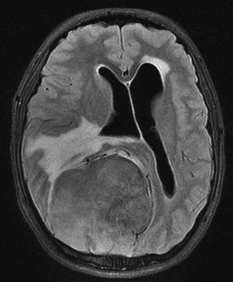
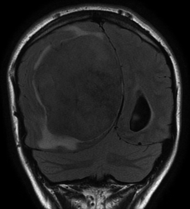
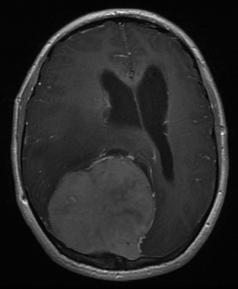
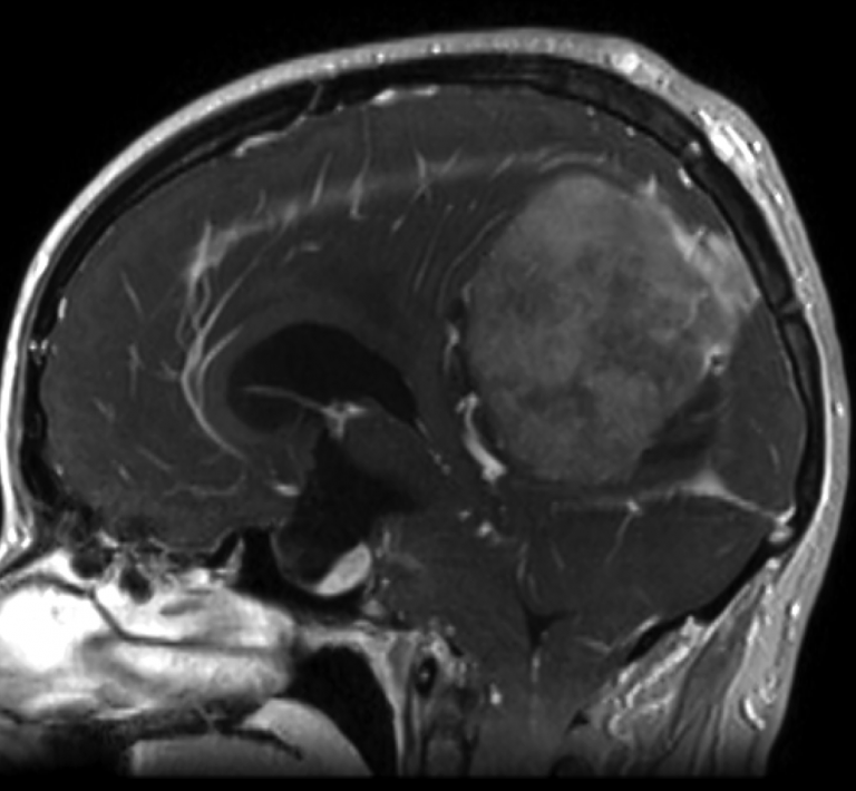
Homogeneously enhancing right parasagittal extra-axial mass with significant mass effect, right-to-left midline shift, obstructive hydrocephalus, and occlusion of the superior sagittal and bilateral transverse sinuses with intracranial to extracranial venous drainage through prominent veins that penetrate the skull.
Differential Diagnosis
Meningioma
Hemangioblastoma
Background
Abnormal communications between the intra- and extra-cranial venous systems are classically termed sinus pericranii.
Sinus pericranii are usually congenital and seen in children. However, these venous anastomoses can rarely be acquired.
Meningiomas commonly demonstrate intravenous extension and dural venous sinus occlusion due to meningiomatous invasion is a well known phenomenon.
In this case, the most likely pathophysiology is that chronic meningiomatous invasion of the intracranial sinuses → venous outlet obstruction → venous hypertension → development of collateral outflow channels.
What's your treatment plan?
There is symptomatic mass effect and obstructive hydrocephalus from this tumor and tumor debulking or resection is warranted.
Cerebrospinal fluid diversion with a ventriculostomy catheter can be useful to reduce intracranial pressure and to achieve brain relaxation.
In this case, because the acquired venous anastomoses, or sinus pericranii are the prominent mechanism of intracranial venous drainage, any surgical treatment of the tumor must carefully protect these venous pathways.
In this case, the entire scalp was carefully shaved and a marking pen and ultrasound were used to outline all prominent scalp veins. This step helped with safe pinning. A frontal ventriculostomy catheter was placed. Image guidance was used to plan a right parasagittal linear incision that totally avoided scalp veins. Craniotomy with near-total tumor resection was performed, and Gamma-Knife radiosurgery was performed in a delayed fashion for the remnant tumor.