Neurovascular Cases

What’s the diagnosis?

The right vertebral artery is diminutive intracranially. The left vertebral artery is larger and dominant. On internal carotid artery injections there is flash filling of the bilateral posterior communicating arteries but they are small. There is a two centimeter mid-basilar artery bilobed irregular aneurysm. It arises between the anterior inferior cerebellar arteries and the superior cerebellar arteries.

3d-reconstruction-x1


Background

  • Aneurysms of the basilar artery trunk are rare and the natural history of these lesions if they go untreated is very poor.
  • These aneurysms are difficult to access using common neurosurgical corridors. Basilar apex aneurysms may be reached using a pterional approach. High basilar artery aneurysms may be reached using a subtemporal approach. And lower, vertebral artery aneurysms may be reached using a far-lateral approach. Pathology at the level of the basilar trunk is anatomically surrounded by the clivus, the brain stem, cranial nerves, and the petrous temporal bones bilaterally. In order to access this “no man’s land” area, skull base approaches that create a window through the petrous temporal bone, called transpetrosal approaches, have been developed.
  • Saccular aneurysms can be clipped at the neck to reconstruct the parent vessel. However, giant, fusiform, atherosclerotic, and dissecting aneurysms may not have an ideal neck across which a clip can be placed. Put differently, there is not always a neck to be occluded.
  • Historically, one microsurgical treatment for such giant aneurysms was ligation of the proximal artery. This technique is called Hunterian ligation. In Hunterian ligation occlusion of the parent artery proximal to the aneurysm → ↓ flow through the aneurysm → aneurysm thrombosis. However, occlusion of carotid or vertebral arteries → ↑ risk of cerebral ischemia.
  • Proximal ligation of the vertebral or basilar arteries has been studied. Similarly, basilar trunk aneurysms have been trapped by placing aneurysm clips proximal and distal to the aneurysm. In such a procedure, flow through robust posterior communicating arteries (PCOMMs) is of critical importance because it may perfuse the posterior cerebral, superior cerebellar, and brainstem perforator territories. Conversely, diminutive PCOMMs → ↑ risk of developing brain-stem ischemia after basilar artery occlusion.

What's your treatment plan?

  • Given the morbidity of open microsurgical approaches, endovascular endoluminal reconstruction of the basilar artery is a reasonable alternative strategy.
  • The Pipeline embolization device is a self-expanding metallic sleeve that adapts to curvy, or tortuous vascular anatomy without kinking or ovalizing.
  • It is made of 48 braided strands of cobalt-chromium and platinum and its metal surface area coverage, or mesh density, is about 30%.

In this case, endovascular remodeling of the basilar artery trunk was performed using the Pipeline embolization device. A total of two telescoping Pipeline devices were placed across the aneurysm. The aneurysm was also partially coil embolized, to further diminish flow into the aneurysm.


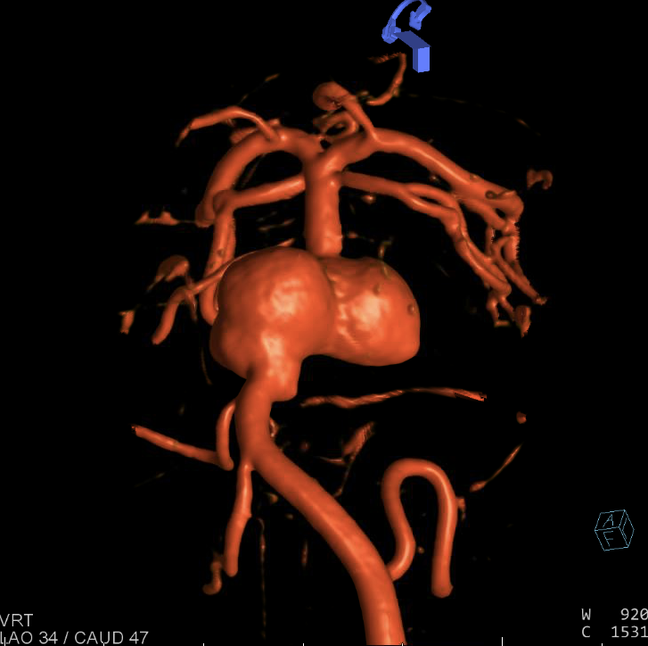
It is thought that initially, the Pipeline device → disruption of flow into the aneurysm. The metallic barrier of the stent redirects blood flow along the course of the parent artery. At the same time, there is ↓ flow into the aneurysm, which → thrombosis within the aneurysm sac.


Over time the device acts as scaffolding over which neointima and endothelium grow and the device becomes incorporated into the vessel wall. Ultimately, this endothelialization process creates a biological seal across the aneurysm.