Neurovascular Cases

What’s the diagnosis?

There are bilateral traumatic epidural hematomas. There is a left sided non-displaced parietal bone fracture with a large convexity epidural hematoma causing left-to-right midline shift. This is an emergent, operative lesion. There is also a right sided non-displaced parietal bone fracture with a smaller epidural hematoma.

post-operative-lateral-skull-x-ray

A left craniotomy was performed with evacuation of the epidural hematoma. Intraoperatively, a lacerated left middle meningeal artery was observed and coagulated using bipolar electrocautery to achieve hemostasis. The bone flap was replaced.


There is significant improvement of the left sided epidural hematoma but residual right sided epidural hematoma.


Differential Diagnosis

  1. There are bilateral skull fractures with associated epidural hematomas in the setting of head trauma. Injury to the bilateral middle meningeal arteries is the most likely diagnosis.

Background

  • Acute epidural hematomas, typically caused by traumatic injury to the middle meningeal artery, commonly enlarge over time and may → acute clinical deterioration.
  • Active hemorrhage from an injured middle meningeal artery is a common cause of hematoma growth, and there are case reports of endovascular embolization of the middle meningeal artery to stop this hemorrhage and prevent hematoma expansion.

What's your treatment plan?

  • In patients with an epidural hematoma who are felt to be threatened to experience hematoma expansion and clinical deterioration, it may be reasonable to consider endovascular embolization of the middle meningeal artery.

In this case, the patient was felt to be at significant risk for expansion of the right sided epidural hematoma, especially in the setting of a recent contralateral hematoma evacuation. Diagnostic cerebral angiography was performed that showed active extravasation from the right middle meningeal artery and endovascular embolization of that vessel was performed to prevent hematoma expansion.

Supra-selective injection of the right middle meningeal artery reveals active extravasation at the area of the cerebrovascular injury. This is conspicuous on both lateral and AP views, and is the target for endovascular embolization.


Post-embolization there is sluggish flow into the proximal middle meningeal artery and no meaningful anterograde flow through the dural component of this vessel. In contrast, the other external carotid artery branches, such as the internal maxillary artery and superficial temporal artery both fill briskly.


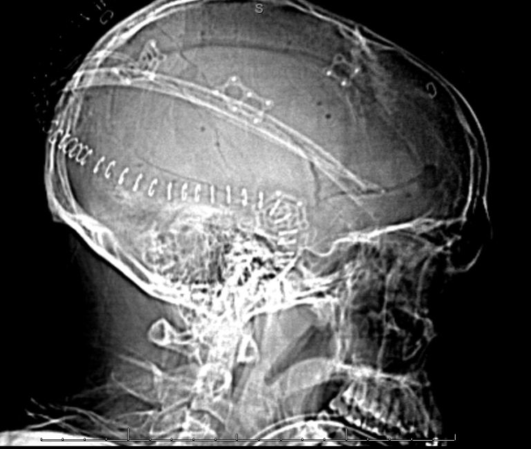
Follow up CT head non contrast several days later demonstrates stability and perhaps even partial resolution of the right-sided epidural hematoma. The previously evacuated left-sided hematoma is also stable.